Leg Bones Diagram - Long Bone Diagram Unlabeled : Humerus Bone Quiz Anterior ... / Key.' carotid canal coronal suture ethmoid.
byAdmin•
0
Leg Bones Diagram - Long Bone Diagram Unlabeled : Humerus Bone Quiz Anterior ... / Key.' carotid canal coronal suture ethmoid.. Posted on january 21, 2015 by admin. File human leg bones labeled svg wikimedia. The largest and most medial leg bone, forming both the knee and ankle joints. Foot bones illustration with icons. Subsequent to the tibia is the fibula, the thinner, weaker bone of the decrease leg.
A leg bone is a bone found in the leg. File human leg bones labeled svg wikimedia. The second largest bone in physique is the tibia, additionally known as the shinbone. Posted on january 21, 2015 by admin. Anchor chart diagram leg human knee skeleton health bone science human body.
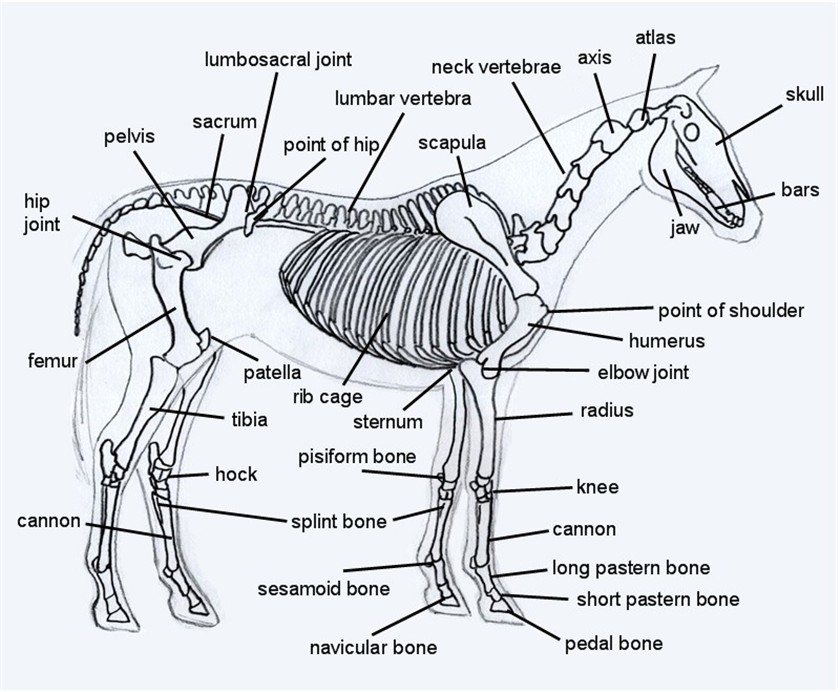
Horse Skeleton Diagram from www.equinespot.com Visit kenhub for more skeletal system quizzes. Subsequent to the tibia is the fibula, the thinner, weaker bone of the decrease leg. Medical diagram with tibia, fibula, malleous, talus and navicular. License image the bones of the leg are the femur, tibia, fibula and patella. Skeletal system label leg diagram quizlet. The bones of the leg are the femur, tibia, fibula and patella. Quizzes on human skeletal system anatomy, bone anatomy, and bone markings. At the same time, the bones and joints of the leg and foot must be strong enough to support the body's weight while remaining flexible enough for movement and balance.
The foot bones shown in this diagram are the talus, navicular, cuneiform, cuboid, metatarsals.
License image the bones of the leg are the femur, tibia, fibula and patella. Joints of hand anterior view, lateral view, right hand. A leg bone is a bone found in the leg. It is also known as the calf bone as it sits slightly behind the tibia on the outside of the leg. Hand | definition, anatomy, bones, diagram, & facts. Posted on april 18, 2019april 18, 2019. File human leg bones labeled svg wikimedia. The foot bones shown in this diagram are the talus, navicular, cuneiform, cuboid, metatarsals. Distal end of right humerus. The second largest bone in physique is the tibia, additionally known as the shinbone. At the same time, the bones and joints of the leg and foot must be strong enough to support the body's weight while remaining flexible enough for movement and balance. These can include any the following: The foot bones shown in this diagram are the talus, navicular, cuneiform, cuboid, metatarsals.
Foot bones illustration with icons. Quizzes on human skeletal system anatomy, bone anatomy, and bone markings. Your leg bones are the longest and strongest bones in your body. File human leg bones labeled svg wikimedia. Top suggestions for human leg bones diagram.
Lower Leg Bones Anatomy - Anatomy Drawing Diagram from fpnotebook.com These muscles work together to produce movements such as standing walking running and jumping. Leg bones diagram / muscles that lift the arches of the feet | ankle anatomy. The largest and most medial leg bone, forming both the knee and ankle joints. Joints of hand anterior view, lateral view, right hand. Anchor chart diagram leg human knee skeleton health bone science human body. The foot bones shown in this diagram are the talus, navicular, cuneiform, cuboid, metatarsals. Bones of the leg and foot, lower leg bone anatomy, leg bones anatomy, leg muscles, leg bones diagram, leg bone structure, leg anatomy muscles, parts of the lower leg. Visit kenhub for more skeletal system quizzes.
Key.' carotid canal coronal suture ethmoid.
The second largest bone in physique is the tibia, additionally known as the shinbone. File human leg bones labeled svg wikimedia. Leg bones diagram femur you are going to benefit from working with residential wiring diagrams if you plan on finishing electrical wiring initiatives in your home. Subsequent to the tibia is the fibula, the thinner, weaker bone of the decrease leg. Most relevant best selling latest uploads. Includes leg (femur, tibia, patella, and fibula) and foot (tarsals and digits) bones. Your leg bones are the longest and strongest bones in your body. Joints of hand anterior view, lateral view, right hand. High resolution textures and displacement included. Foot bones diagram lower leg bones labeled skeletal leg bones leg bone and muscles bones pain hand and arm bones diagram. The foot bones shown in this diagram are the talus, navicular, cuneiform, cuboid, metatarsals. Quizzes on human skeletal system anatomy, bone anatomy, and bone markings. Human anatomy diagrams show internal.
These muscles work together to produce movements such as standing walking running and jumping. Your leg bones are the longest and strongest bones in your body. Key.' carotid canal coronal suture ethmoid. File human leg bones labeled svg wikimedia. Skeleton leg ankle joints and toe.
human leg bone anatomy | Human leg, Leg bones, Anatomy from i.pinimg.com Foot bones illustration with icons. Quizzes on human skeletal system anatomy, bone anatomy, and bone markings. Includes leg (femur, tibia, patella, and fibula) and foot (tarsals and digits) bones. Key.' carotid canal coronal suture ethmoid. It is also known as the calf bone as it sits slightly behind the tibia on the outside of the leg. Human anatomy diagrams show internal. These can include any the following: The foot bones shown in this diagram are the talus, navicular, cuneiform, cuboid, metatarsals and calcaneus.
High quality realistic skeleton legs.
Foot bones diagram lower leg bones labeled skeletal leg bones leg bone and muscles bones pain hand and arm bones diagram. These can include any the following: Your leg bones are the longest and strongest bones in your body. Medical diagram with tibia, fibula, malleous, talus and navicular. High quality realistic skeleton legs. These muscles work together to produce movements such as standing walking running and jumping. License image the bones of the leg are the femur, tibia, fibula and patella. Posted on january 21, 2015 by admin. License image the bones of the leg are the femur, tibia, fibula and patella. Skeletal system label leg diagram quizlet. The bones of the leg are the femur, tibia, fibula and patella. Visit kenhub for more skeletal system quizzes. Subsequent to the tibia is the fibula, the thinner, weaker bone of the decrease leg.